
• Add description, images, menus and links to your mega menu

  • A column with no settings can be used as a spacer

  • Link to your collections, sales and even external links

  • Add up to five columns

  • da

  • A column with no settings can be used as a spacer

  • Link to your collections, sales and even external links

  • Add up to five columns

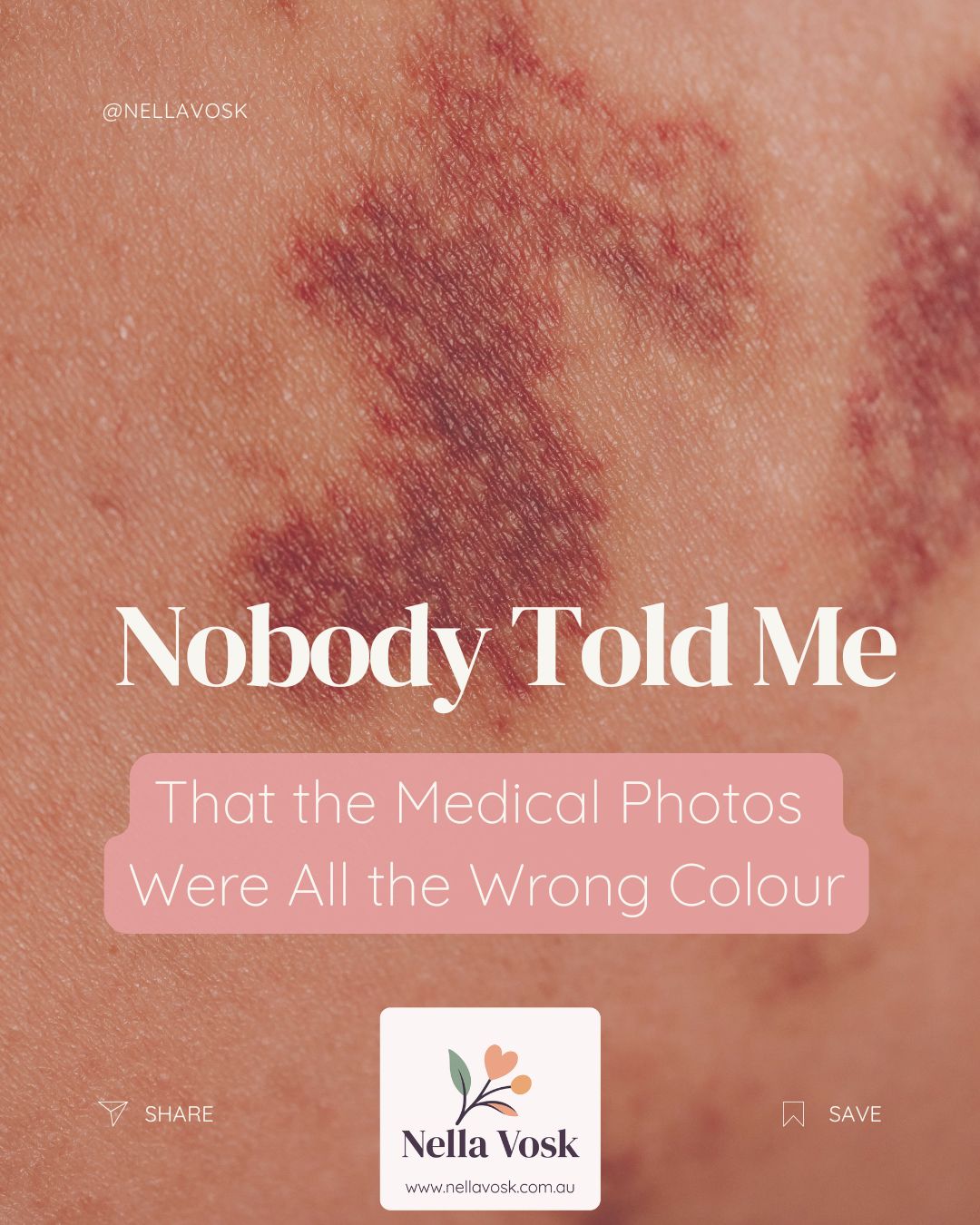
  • Nobody Told Me That the Medical Photos Were All the Wrong Colour

    Founder of Nella Vosk • 14+ years supporting families across motherhood, feeding, and early childhood wellbeing

    Nobody Told Me That the Medical Photos Were All the Wrong Colour

    This article is part of the Nella Vosk Complete Guide to Baby Eczema for Australian Families — everything you need to know about managing eczema-prone skin from the early months through toddlerhood.

    This article is part of Nella Vosk’s Nobody Told Me series — a growing collection of posts written for the parent who has been trying everything and still feels like they’re failing their child. Every post in this series starts from a moment a parent wasn’t given information they needed, and works back through what the evidence actually says. You can find the full series at nellavosk.com.au/pages/nobody-told-me.


    Nobody told me that the medical photos were all the wrong colour.

    When my child’s skin started changing — patches that were darker than the surrounding skin, areas that looked grey or purple-tinged, a loss of colour in places where eczema had previously been active — I searched for images to compare. Everything I found showed the same thing: red, inflamed patches on light skin. I couldn’t find a single image that looked like my child’s skin. I assumed I was dealing with something different. Something more serious. Something the eczema resources weren’t covering.

    What I wasn’t told was that eczema on brown, dark, or deeply pigmented skin simply does not look like eczema on light skin. The redness that is the signature visual feature of atopic dermatitis in most published images, textbooks, and training materials is a function of dilated blood vessels visible through pale skin. In darker skin tones, the same underlying inflammation often appears grey, purple, dark brown, or ashen rather than red. The itch is the same. The skin barrier dysfunction is the same. The treatment is the same. But the appearance is so different that it is frequently missed by GPs, maternal and child health nurses, and parents themselves — because nobody told them what to look for.

    This post explains how eczema looks across different skin tones, why it changes the colour of the skin, whether the colour comes back, and what it means for getting a correct and timely diagnosis in Australia.

    The Diagnosis Gap: Why Eczema Is Missed on Darker Skin

    The underdiagnosis of eczema — and of inflammatory skin conditions generally — in people with darker skin tones is a documented clinical problem with a documented structural cause: most of the published dermatological images that clinicians are trained on, that textbooks illustrate, and that diagnostic reference materials use depict conditions on white or light skin.

    Research published in the journal Clinical and Experimental Dermatology found that the majority of clinical images used in dermatology education across English-speaking countries depicted conditions on Fitzpatrick skin types I to III (light skin), with minimal representation of skin types IV to VI (medium, brown, and dark skin). A separate study examining medical student diagnostic accuracy found that students were significantly more likely to correctly diagnose eczema on lighter skin than on darker skin — even when the underlying clinical condition was identical. The diagnostic confidence gap was consistent and statistically significant.

    In Australia, where approximately 30% of the population identifies as having Asian, Middle Eastern, African, Pacific Islander, or First Nations heritage, this diagnostic gap has real clinical consequences. Children with eczema who have darker skin are more likely to receive delayed diagnosis, more likely to be misdiagnosed with other conditions, and more likely to have their condition underestimated in severity because the visual reference points clinicians use are calibrated for lighter skin.

    This is not a criticism of individual clinicians. It is a systemic training problem. But for a parent navigating their child’s eczema, it means that self-education about what eczema looks like on their child’s specific skin tone is not just useful — it may be necessary for getting the correct diagnosis in the first place.

    What Does Eczema Look Like on Darker Skin?

    The visual presentation of eczema varies significantly across the skin tone spectrum. Understanding these differences is the first step to recognising the condition in a child with brown or dark skin.

    Colour of inflamed areas

    In light skin (Fitzpatrick types I-II): the classic “red and inflamed” appearance. Dilated blood vessels are highly visible through pale skin, producing the red, pink, or scarlet patches that most eczema images show.

    In medium and olive skin (Fitzpatrick types III-IV): inflammation may appear as a dusky red, brownish-red, or darker brown discolouration against the surrounding skin. True redness may still be present but is less pronounced and harder to identify.

    In brown and dark skin (Fitzpatrick types V-VI): inflammation most commonly appears as a grey, purple, violet, or dark brown discolouration. The skin may look ashen or dull in affected areas. The characteristic redness of eczema on light skin may be barely visible or completely absent. Patches may appear darker rather than redder than surrounding skin during active inflammation.

    Can eczema go purple? Yes — on darker skin tones, purple or violet-toned patches are a common presentation of active eczema inflammation. This is not a sign of something more serious; it is the same inflammatory process visible differently through more melanin-rich skin. If your child has darker skin and their eczema patches appear purple, grey, or violet, this is within the typical presentation range for their skin tone.

    Texture and scaling

    Across all skin tones, eczema produces textural changes that are more consistent than colour changes: dry, rough, or thickened patches; scaling or flaking; lichenification (a leathery thickening of the skin that develops with chronic scratching); and the characteristic location patterns of atopic dermatitis (face and scalp in infants; elbow creases, knee creases, and neck in older children). These textural signs are visible on all skin tones and are often the more reliable diagnostic indicators when colour presentation is ambiguous.

    Location patterns

    The distribution of eczema in babies and young children is consistent across skin tones: cheeks, forehead, and scalp in early infancy; gradually moving to the flexural creases (inner elbows, behind knees, wrists, ankles) in toddlerhood. Eczema in these locations on a child with dark skin may not look red but will still itch, still feel rough or dry, and still follow the same flare-and-remission pattern.

    Follicular prominence

    In children with darker skin, eczema may present predominantly as follicular eczema — a pattern in which the inflammation centres on the hair follicles, producing a bumpy, rough texture (sometimes described as “goosebump-like”) rather than the flat, scaly patches more typical on lighter skin. This pattern is commonly mistaken for keratosis pilaris or simple dry skin, and the eczema component is missed.

    Why Does Eczema Change Skin Colour?

    The skin colour changes associated with eczema — both during and after active inflammation — occur through several distinct mechanisms:

    Active inflammation: vasodilation

    During an active eczema flare, the immune response dilates blood vessels in the skin (vasodilation) and increases blood flow to the affected area. In light skin, this is visible as redness. In darker skin, the increased melanin concentration in the epidermis partially masks the red colour of the underlying vasodilation, producing the grey, purple, or brown discolouration described above.

    Post-inflammatory hyperpigmentation (PIH)

    After an eczema flare resolves — whether through treatment or natural remission — the affected skin often temporarily darkens. This is called post-inflammatory hyperpigmentation. The inflammatory process triggers increased melanin production in the epidermis, and the increased pigmentation persists for weeks to months after the inflammation has cleared. PIH is significantly more common and more pronounced in people with darker skin tones, because higher baseline melanin density means the inflammatory pigmentation signal is amplified.

    PIH presents as flat, dark brown or greyish-brown patches at the site of previous eczema. The patches are not inflamed, not itchy (or minimally so), and not scaly — they are simply darker than the surrounding skin. They are not a sign that the eczema is still active. They are the skin’s response to the previous inflammation.

    Post-inflammatory hypopigmentation

    In some children with darker skin, eczema can also produce post-inflammatory hypopigmentation — patches that are lighter than the surrounding skin after a flare. This occurs when the inflammatory process disrupts melanocyte function (the cells responsible for producing melanin), temporarily reducing pigment production in the affected area. Hypopigmented patches can be alarming for parents who have not been told to expect them, as they can resemble vitiligo or other depigmentation conditions.

    Both post-inflammatory hyperpigmentation and post-inflammatory hypopigmentation are temporary in most cases. They resolve as the skin recovers and normal melanocyte activity is restored, though this can take months.

    Will My Child’s Skin Colour Come Back After Eczema?

    Yes — in the vast majority of cases, the skin colour changes caused by eczema are temporary and resolve as the skin heals.

    • Post-inflammatory hyperpigmentation typically fades over weeks to months as new skin cells replace the hyperpigmented cells. In children, whose skin turns over more rapidly than adult skin, this process can be faster. Consistent use of sun protection on affected areas can help by preventing UV from further stimulating melanin production in already-hyperpigmented patches

    • Post-inflammatory hypopigmentation also typically resolves as melanocyte function recovers. The recovery timeline varies but is usually complete within three to twelve months of the eczema flare resolving

    • The most important factor in resolving post-inflammatory colour changes is controlling the underlying eczema. Each new flare resets the skin’s recovery timeline. Consistent eczema management — daily emollient use, prompt treatment of flares, trigger reduction — gives the skin the stable periods it needs to recover both its barrier function and its pigmentation

    If your child has colour changes on their skin that you cannot confidently attribute to their eczema history, or if the discolouration appears to be spreading, worsening, or not resolving over time, a GP assessment is appropriate. Post-inflammatory pigmentation changes are common and expected, but unusual pigmentation warrants professional assessment to rule out other conditions.

    How to Advocate for Your Child When the Visual Doesn’t Match the Textbook

    The diagnostic gap in eczema on darker skin means that some parents will need to do more than present their child to a clinician and wait for a diagnosis. Here is what helps:

    • Describe the symptoms, not just the appearance. Itch intensity (particularly overnight scratching), skin roughness, flare patterns, distribution, and the child’s sleep disruption are consistent across skin tones and give a clinician useful diagnostic information even when the visual presentation is atypical

    • Photograph the skin at its worst. Eczema is cyclical, and a GP appointment may not coincide with an active flare. Photographs taken during flares, including close-up images of texture as well as colour, give the clinician a more complete picture than an in-consultation view of recovering skin

    • Name the diagnosis you are considering. It is appropriate to say: “I have been reading about eczema and I am concerned this is what my child has. I’m aware that eczema can look different on darker skin. Can we discuss whether this fits?” Most GPs will respond constructively to a parent who has clearly done their research

    • Request a referral to a paediatric dermatologist if the eczema is not responding to first-line GP management, or if the diagnosis remains uncertain. A paediatric dermatologist has specialist training in recognising eczema across skin tones and is the most appropriate referral pathway for complex or treatment-resistant cases

    The Treatment Is the Same — Regardless of Skin Tone

    One of the most important things to know is that once eczema is correctly identified, the treatment principles are identical across all skin tones. The emollient routine, the three-minute window, the topical corticosteroid sequence, the trigger identification, the bedding hygiene, the scratch protection at night — all of these work the same way regardless of whether the eczema presents red, purple, grey, or brown.

    The NTM series posts that cover these management approaches apply to every family managing eczema, regardless of their child’s skin tone:

    Nobody Told Me That the Order I Applied the Skincare Mattered — the three-minute window and skincare sequence

    Nobody Told Me My Baby Was Scratching All Night Without Even Waking Up — the overnight itch-scratch cycle

    Nobody Told Me My Baby’s Eczema Wasn’t My Fault — the genetic basis and cause-vs-trigger framework

    Keep Reading

    Infected Eczema in Babies: Signs, Symptoms & When to Act

    Why Kids Scratch More at Night: The Physiology Explained

    How to Stop Baby Scratching at Night Without Overdressing Them

    Eczema Mittens vs Sleeves: Which Is Better?

    Wet Wrapping for Eczema: How It Works & When to Use It


    Eczema management works the same way regardless of skin tone. What protects the skin barrier overnight — breathable fabric, integrated scratch protection, thermoregulation — works the same way for every child. The Bamboo Bubby range was built for the child with eczema, full stop. Every skin tone. Every presentation. Every night.


    ABOUT THE AUTHOR

    Kelly Northey is a Certified Postpartum Nutrition Professional (CPPNP) and founder of Nella Vosk, the parent company of the Bamboo Bubby range. She has spent fourteen years working with Australian families navigating baby and child eczema — the sleepless nights, the skin that won’t heal, and the guilt of feeling like you should be doing more. The Bamboo Bubby range exists because of those families.
    Learn more about Kelly.


    Also in Nobody Told Me: Your Baby's Skin

    Nobody Told Me I Had to Fight That Hard Just to See a Dermatologist
    Nobody Told Me I Had to Fight That Hard Just to See a Dermatologist

    Read More
    Nobody Told Me That the Antibiotics for the Infected Eczema Might Make the Next Infection More Likely
    Nobody Told Me That the Antibiotics for the Infected Eczema Might Make the Next Infection More Likely

    Read More
    Nobody Told Me That Adult Eczema Needs Different Management — or That My Baby’s Approach Wouldn’t Work for Mine
    Nobody Told Me That Adult Eczema Needs Different Management — or That My Baby’s Approach Wouldn’t Work for Mine

    Read More